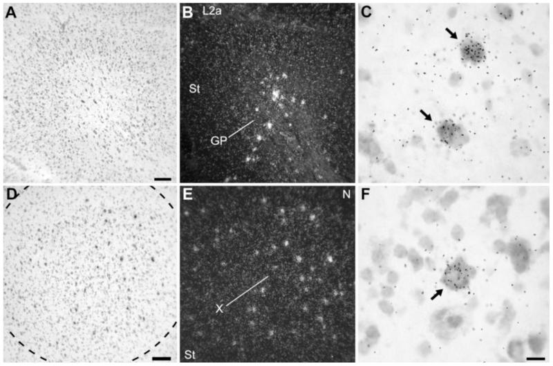
Fig. 9.
NF-M expression in the basal ganglia. A,B: Brightfield view of Nissl staining and darkfield view of emulsion autoradiography of parasagittal section depicting the caudal globus pallidus (GP), at about the same level as in Figure 2C. C: High-magnification view of labeled cells in GP. Arrows point to the sparse large neuronal cells with high accumulation of emulsion grains. D,E: Brightfield view of Nissl staining and darkfield view of emulsion autoradiography of frontal section through song nucleus area X in the medial striatum. F: High-magnification view of labeled cells within area X. Arrows point to widely spaced large neuronal cells with high accumulation of emulsion grains. For A–C, dorsal is upward and rostral is to the right; for D–F, dorsal is upward and lateral is to the right. For abbreviations see list. Scale bars = 100 μm in A (applies to A,B); 100 μm in D (applies to D,E); 20 μm in F (applies to C,F).

