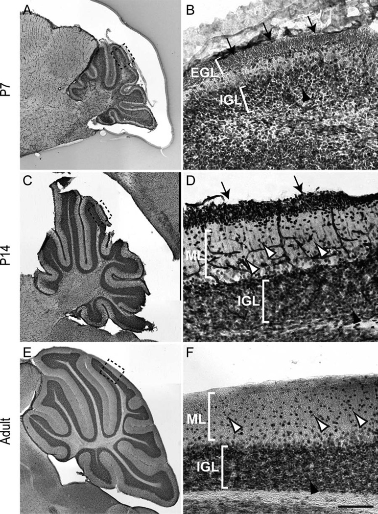
Figure 1.
Expression of NFIX in the cerebellum. Expression of NFIX in sagittal sections of the postnatal (A–D) and adult (E,F) cerebellum. A: NFIX was broadly expressed in the cerebellum at P7. B: Higher magnification view of the boxed region (lobule VI) in A, showing expression of NFIX by cells within the external granular layer (EGL; arrows) and inner granule cell layer (IGL; arrowhead). C: Expression of NFIX at P14. D: Higher magnification view of the boxed region (lobule VI) in C, showing that immature granule neurons within the premigratory zone of the EGL (arrows) and cells within the molecular layer (ML; open arrowheads) and IGL (solid arrowhead) express NFIX. E: Expression of NFIX in the adult cerebellum. F: Higher magnification of the boxed region (lobule VI) in E. NFIX was expressed by cells within the IGL (solid arrowhead), as well as by scattered cells within the ML (open arrowheads). Scale bar = 25 µm in F (applies to D,F); 200 µm for A,C,E; 15 µm for B.

