Figure 3.
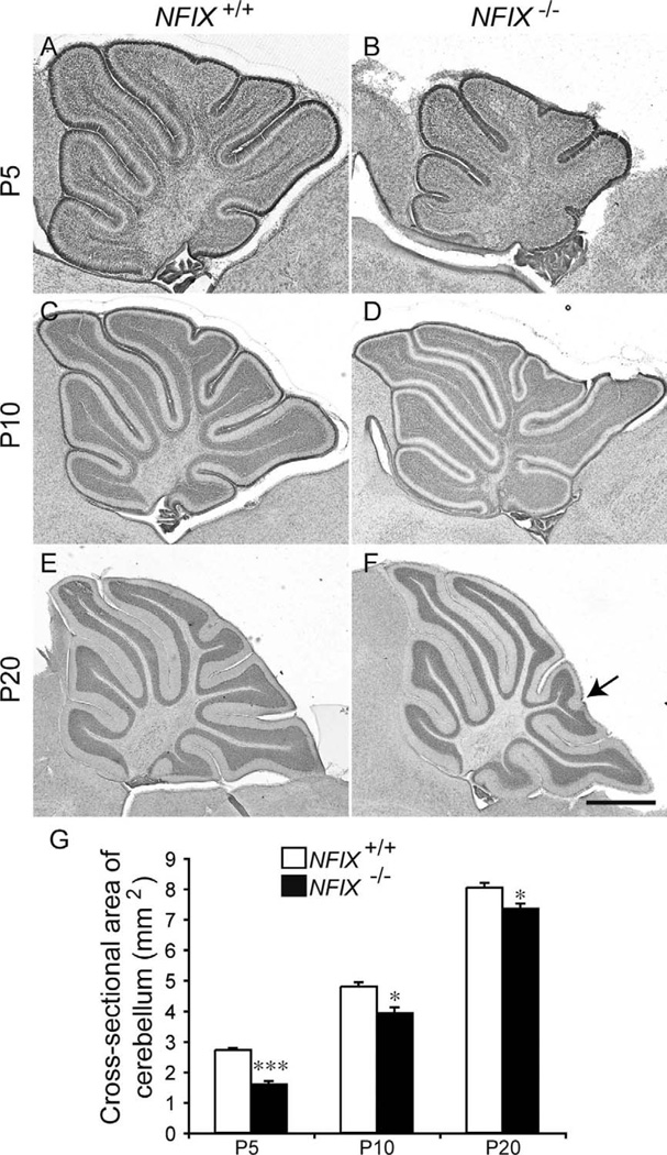
Reduction in cerebellum size in NFIX−/− mice. Hematoxylin-stained sagittal sections of the cerebellum of wild-type (A,C,E) and NFIX−/− (B,D,F) mice at P5 (A,B), P10 (C,D), and P20 (E,F). G: Quantification of the area of the cerebellum at the level of the vermis. At P5, the cross-sectional area of the NFIX−/− cerebellum (B) was markedly smaller than that of the wild-type (A). At P10 and P20 the size of the NFIX−/− cerebellum (D,F) had partially recovered but was still significantly reduced in comparison with that of wild-type controls (C,E). The folia of NFIX−/− mice appeared subtly different at P20; for instance, the tip of lobule VIII appears bifurcated in the mutant (arrow in F). *P < 0.05; ***P < 0.001, t-test. Scale bar = 200 µm in F (applies to E,F); 100 µm for A,B; 150 µm for C,D.

