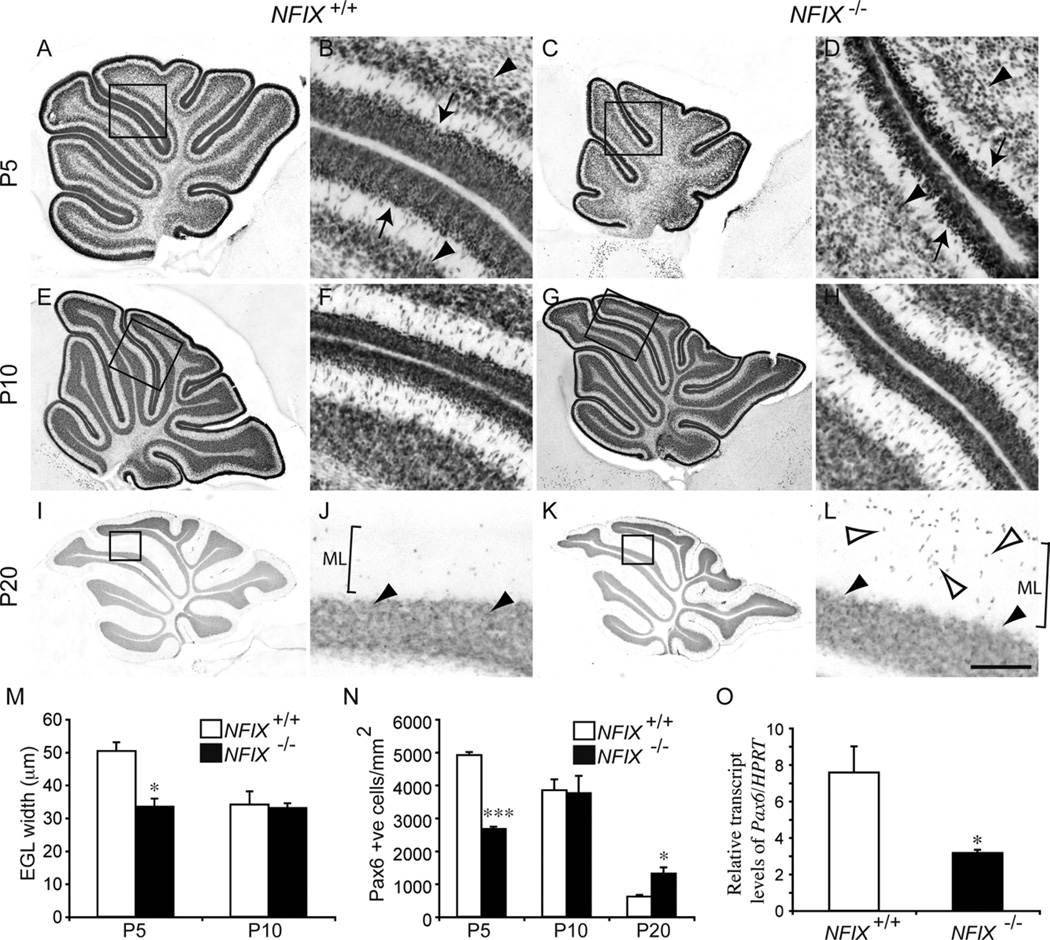
Figure 5.
Delayed differentiation of the EGL layer in NFIX−/− mice. Expression of Pax6 at P5 (A–D), P10 (E–H), and P20 (I–L) in wild-type and NFIX−/− mice. A: Expression of Pax6 in the wild-type cerebellum at P5. B: Higher magnification of the boxed region in A (lobules IV– VI), revealing that cells within the EGL (arrows) and the nascent IGL (arrowheads) express Pax6. C: Expression of Pax6 in the NFIX−/− cerebellum at P5. D: Higher magnification of the boxed region in C (lobules IV–VI), revealing that the width of the EGL was markedly reduced (arrows) and that the IGL (arrowheads) was less well defined. By P10, expression of Pax6 in the cerebellum of wild-type (E,F) and NFIX−/− mice was comparable. By P20, the EGL was no longer evident in either the wild-type (I,J) or the NFIX−/− (K,L) cerebella. Cells within the IGL maintained Pax6 expression (solid arrowheads in J,L). However, in the mutant cerebellum, there were markedly more Pax6-positive cells in the molecular layer (open arrowheads in L) than were observed in the control. F,H,J,L are higher magnification views of the boxed regions in E,G,I,K, respectively (lobules IV–VI in F,H; lobules IV/V in J,L). M: The width of the EGL was significantly reduced in the cerebellum of NFIX−/− mice at P5 compared with controls. N: Counts of Pax6-positive cells in the molecular layer of wild-type and mutant cerebella. At P5 there were significantly fewer Pax6-positive cells in the molecular layer of NFIX−/− mice. However, at P20 there were significantly more Pax6-positive cells in the molecular layer of the mutant compared with wild-type controls. O: qPCR on P5 cerebellar tissue revealed a significant reduction in Pax6 mRNA in the NFIX−/− mutant compared with controls. ML, molecular layer. *P < 0.05; ***P < 0.001, t-test. Scale bar = 40 µm in L (applies to B,D,F,H,J,L); 100 µm for A,C; 150 µm for E,G; 200 µm for I,K.

