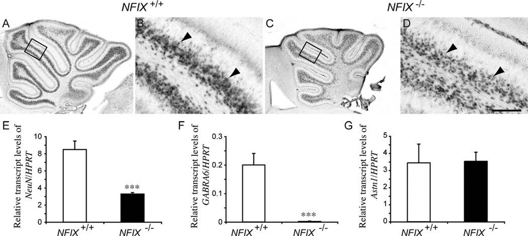
Figure 7.
Neuronal differentiation is delayed in the cerebellum of NFIX−/− mice. Expression of the neuronal marker NeuN in the cerebellum of wild-type (A,B) and NFIX−/− (C,D) mice at P5. NeuN-expressing cells in the IGL of the wild-type and mutant were apparent (arrowheads), but there appeared to be more NeuN-positive cells in the IGL of the wild-type compared with the NFIX knockout. B,D are higher magnification views of the boxed regions (lobules IV/V) in A,C, respectively. E–G: qPCR on wild-type and NFIX−/− P5 cerebellar tissue demonstrated significantly reduced levels of NeuN (E) and GABRA6 (F) mRNA in the mutant. However, levels of astrotactin 1 mRNA (G) were not significantly different compared with controls. ***P < 0.001, t-test. Scale bar = 20 µm in D (applies to B,D); 100 µm for A,C.

