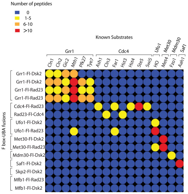
Figure 2. LC-MS/MS analysis of two-step purifications of Ligase Traps identifies known SCF substrates.
Color-coded matrix showing known SCF substrates identified by Ligase Trapping. Two-step purifications were performed from cell extracts expressing UBA fusions of eight F-box proteins. These represent data from initial purifications performed in parallel. In the case of Cdc4, both N- and C-terminal purifications are shown. Dsk2 and Rad23 fusions are shown when both fusions were well expressed. Repeats of two identical pairs of Grr1 traps are shown for comparison. Colors represent spectral counts for each protein in each purification. For full list of substrates and references, see Table S1.

