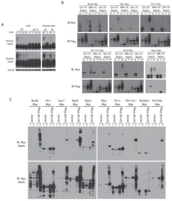
Figure 4. Ubiquitination of Grr1 candidate substrates.
(A) Short and long exposures of anti-Myc Western blots of a CHX chase assay of the uncharacterized ORF YKR045C in GRR1 and grr1Δ cells. Steady state levels of Ykr045c are shown in G1- and nocodazole-arrested cells. (B) Six candidate Grr1 substrates were expressed in cells containing Ligase Traps of Grr1 as well as Mfb1 and/or Ufo1 as negative controls. Western blots of two-step purifications, as in Figure 1D, are shown. (C) Western blots of whole cell extract (I) and anti-Flag pulldowns (P) from strains expressing Myc-tagged candidate Grr1 substrates and transformed with either empty vector (pRS426) or pYES2-Grr1ΔF-Fl (a galactose-inducible copy of Grr1-Flag lacking the F-box domain).

