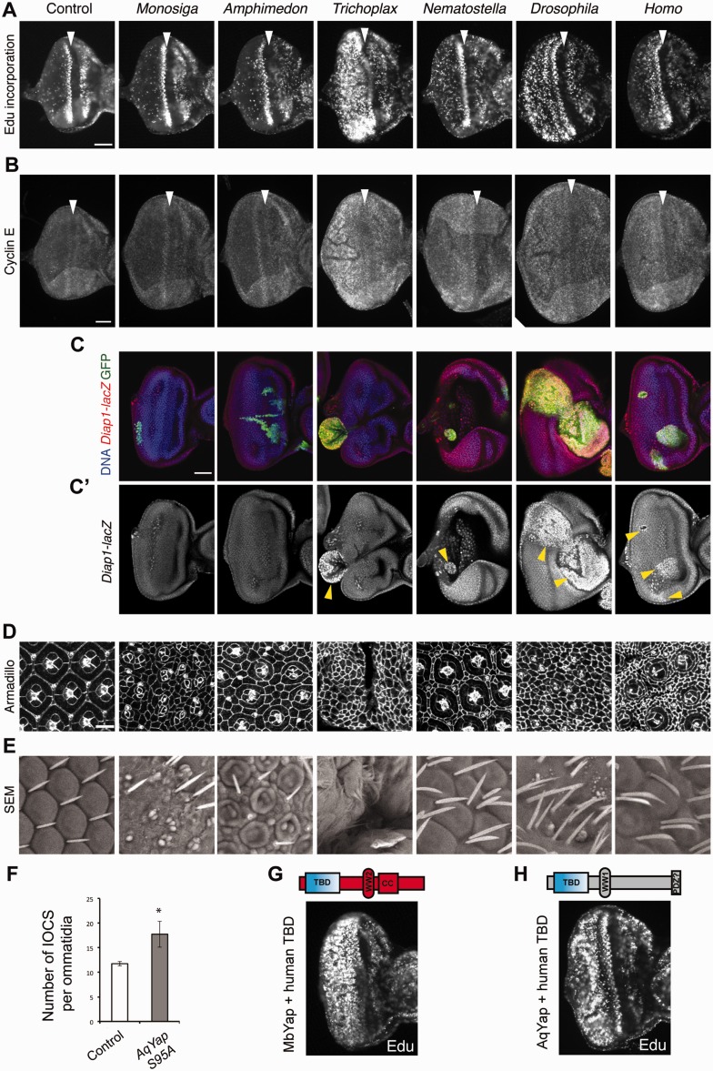
Fig. 3.
Cellular and molecular mechanisms underlying Yap ortholog-induced overgrowth in Drosophila. (A) Edu labeling in eye discs overexpressing hypothetically nonphosphorylatable forms of Yap from the indicated species under the control of GMR-Gal4. Note the dramatic induction of proliferation by Trichoplax yap and the absence of ectopic proliferation caused by the Monosiga and Amphimedon orthologs. (B) Immunostaining of Cyclin E in eye discs of the same genotypes indicated above. Arrowheads indicate the position of the morphogenetic furrow. Scale bar = 50 μm. (C-C′) diap1-lacZ expression (red) in eye discs overexpressing the corresponding Yap orthologs in clones (GFP+, green) and stained with Hoechst (blue). Except for AqYapS95A and MbYapS48A, elevated diap1-lacZ expression was detected in all Yap ortholog-overexpressing clones (yellow arrowheads). Scale bar = 50 μm. (D) Pupal retinae from the genotypes indicated above, stained with anti-Armadillo antibody to visualize cell outlines at 42 h after puparium formation. Scale bar = 10 μm. (E) Corresponding scanning electron micrographs (SEM) of adult retinae. (F) Quantification of IOCs per ommatidia in controls (w, GMR-Gal4/+; n = 20) and following expression of Amphimedon Yap (GMR-Gal4>AqYapS95A; n = 20). Statistical analysis was performed using Student’s t-test (*P < 0.001). (G, H) Edu incorporation assay in eye discs overexpressing chimeric constructs of Monosiga Yap (G) and Amphimedon Yap (H) with the Homo TBD. Addition of the human TBD to either variant resulted in a strong capacity to induce proliferation.

