Fig. 5.
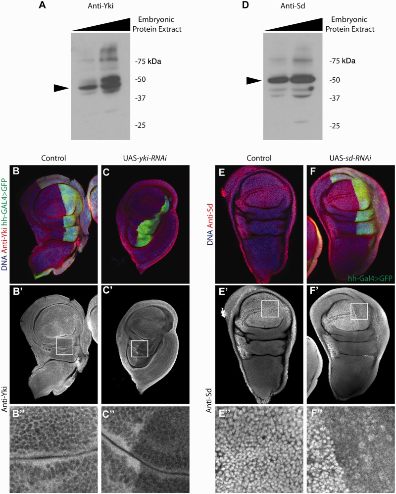
Validation of anti-Yki and anti-Sd antibodies. (A) Western blot analysis shows that anti-Yki antibody detects a strong signal at the expected molecular weight (arrowhead, 45 kDa). (B-B″) Control wing disc stained with Hoechst (blue) and anti-Yki (red). The posterior compartment is marked by the expression of hh-Gal4>UAS-GFP (green). Anti-Yki detects ubiquitous expression of Yki. (C-C″) Wing disc overexpressing UAS-yki-RNAi under the control of hh-Gal4. The clear reduction of anti-Yki staining in the posterior compartment (inset box) confirms that our antibody recognizes Drosophila Yki. (D) Anti-Sd detects a specific band at the expected molecular weight (arrowhead, 50 kDa). (E-E″) Control wing disc stained with Hoechst (blue) and anti-Sd (red). Endogenous sd expression is elevated in the wing pouch and margin, which is consistent with our Anti-Sd staining. (F-F″) A wing disc expressing UAS-sd-RNAi under the control of hh-Gal4 shows a strong reduction of Sd staining in the posterior compartment (inset box), confirming the specificity of Anti-Sd.

