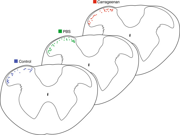
Figure 2.

Location of recorded neurons in spinal cord slices. The location of recorded neurons was documented to check and account for bias. Low magnification images (5×) were captured at the conclusion of recording sessions with the recording pipette still in place. Recording locations were then plotted on a standardised transverse template of the upper cervical spinal cord. Recording locations were then consolidated onto a single template for control, sham, and carrageenan-injected experiments. Comparison of these maps indicates similar sampling in both mediolateral and dorsoventral planes for each experimental condition.
