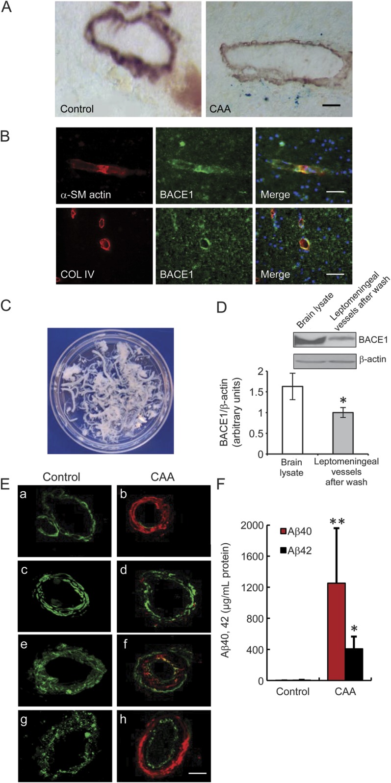
Figure 1. BACE1 expression in human cortical and leptomeningeal vessels.

(A) Microbleeds were found in the brains of patients with CAA. The small arteries were identified with anti–α-SM actin, a smooth muscle cell marker. Cerebral microbleeds were identified by presence of hemosiderin (iron), using Prussian blue stain. A staining of ferric irons (blue) was observed in α-SM actin–positive vessels in the frontal cortex of patients with CAA, whereas it was not observed in the brains of controls without dementia. Bar: 50 μm. (B) BACE1 expression in the human cortical vessels marked by α-SM actin and collagen IV antibodies. Bar: 50 μm. (C) Leptomeninges were dissected from the brain surface in the immediate postmortem period and then each isolated vessel was washed with saline on ice until no single red blood cell could be observed, as seen in white color. (D) BACE1 expression in different tissues, including human frontal cortex and leptomeningeal vessels after wash, to confirm the expression of BACE1 in leptomeninges (*p < 0.05). (E) Vascular amyloid deposits and vascular degeneration in the leptomeningeal vessels of subjects with CAA and controls. A significant Aβ40 (red) deposition and loss of smooth muscle cells (α-SM actin, green) were observed in the leptomeningeal vessels of subjects with CAA (a, b). Aβ42 (red) deposition and loss of smooth muscle cells (α-SM actin, green) were observed in the leptomeningeal vessels of subjects with CAA (c, d). A significant Aβ (red) deposition was observed in the leptomeningeal vessels of subjects with CAA, which was labeled with antibody against the vessel basement membrane collagen IV (e, f, green), as well as the degeneration of endothelial cells, which was marked by antibody against von Willebrand factor (g, h, green). Bars: 20 μm. (F) Quantitative determination of Aβ40 and Aβ42 in the leptomeningeal vessels of patients with CAA and controls by ELISA assay. The leptomeningeal vessels of patients with CAA showed a significantly increased amount of Aβ40 and Aβ42 proteins compared with those of controls, which confirmed that the investigated leptomeningeal vessel samples showed the presence of CAA (*p < 0.05, **p < 0.01). Error bars represent SD. Aβ = β-amyloid; α-SM = α-smooth muscle; BACE1 = β-site APP-cleaving enzyme 1; CAA = cerebral amyloid angiopathy; COL = collagen.
