
Figure 4. Immunohistochemical Analysis of Transporter Gene Expression in the Retina.
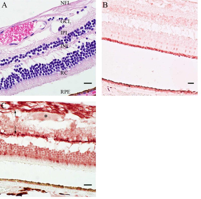
Control retina sections were prepared with hematoxylin-eosin (H & E) staining (A) to differentiate the tissue layers. The protein expression pattern of two transporters was evaluated by IHC: OCT3 (B) and BCRP (C). (C) The arrow (↑) and the asterisk (*) denote nerve fibers and a blood vessel, respectively. Scale bars are set to 20 microns. See Supplemental Table S1 for donor information. An eye pathologist reviewed all IHC of the retina. RPE, retinal pigmented epithelium; RC, rods and cones; INL, inner nuclear layer; IPL, inner plexiform layer; GCL, ganglion cell layer; NFL, nerve fiber layer.