
Figure 3. 2D electrophoretic profiling of Suberites domuncula primmorph extracts in response to 10−6 M of N-3-oxododecanoyl-L-homoserine lactone (3-oxo-C12-HSL).
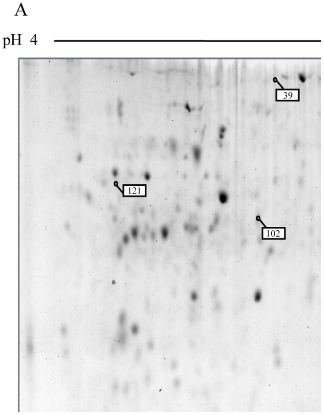
Images correspond to representative gels of a total 3-oxo-C12-HSL-stimulated protein primmorph extract separated with a linear gradient pH 4–7 (A) or pH 5–8 (B), and of a membrane 3-oxo-C12-HSL-stimulated protein primmorph extract separated with a linear gradient pH 5–8 (C). Spots with amounts significantly (p<0.05) changed are highlighted on the gels with white and black boxes for protein, the amount of which is significantly higher or lower in presence of 3-oxo-C12-HSL, respectively. Enlargements of these spots are shown in D (left part: 3-oxo-C12-HSL -treated sample; right part: untreated sample).