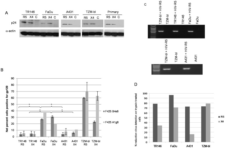
Figure 3. Different methods used to detect HIV-1 R5 and X4 binding to epithelial cells.
(A) Post-lysis detection of p24 gag protein by Western blotting. Primary (gingival) epithelial cells, TR146, FaDu, A431 and TZM-bl cells were incubated overnight (16–24 h) with cell free YU2 (R5) or LAI (X4). After extensive washing to remove unbound virus, normalised total protein lysates were separated by SDS-PAGE and probed for HIV p24 using α-actin as a loading control. (B) Detection of immobilized virus on the cell surface by flow cytometry. Epithelial cells were incubated overnight with cell free virus. Bound virus was detected using a Cy5-labeled anti-human secondary antibody to detect HIV-1 gp120 primary monoclonal on the APC channel. Electronic gates were set around an unlabelled cell control, this area is then set as zero and any cells shifted to the right of the gate are deemed positive. To determine amount of virus bound, virally exposed, labelled cell percentages are subtracted from the uninfected (unexposed) labelled control cell percentages to obtain the % fluorescence values shown. Data are representative of four independent experiments and bars indicate ± standard deviation from the mean. (C) Detection of packaged HIV R5 RNA by amplification of the HIV-1 pol gene using nested PCR. Total RNA was extracted from TR146, FaDu, A431 and TZM-bl cells incubated overnight with cell free YU2 (R5) and used to produce viral cDNA. This was then used as a template in a nested PCR to detect a 2 Kb region of HIV pol. (D) Percentage reduction in detection of immobilized virus on the cell surface by flow cytometry after trypsin treatment. Virally exposed cells are compared with cells labelled with secondary antibody alone. Data set is representative of three independent experiments. * = P<0.05.

