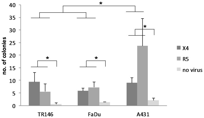
Figure 7. Transfer of captured HIV-1 from epithelial cells to permissive cells via transcytosis.
TR146, FaDu and A431 were grown on polycarbonate transwell membranes and incubated with R5 (YU2) and LAI (X4) virus for 4 h. Following extensive washing the transwells were placed in a separate plate that overlaid a confluent monolayer of TZM-bl cells and incubated for a further 48 h at 37°C. TZM-bl cells were fixed, stained for β-galactosidase expression with X-Gal stain, and counted using light microscope at 100x magnification. Resulting replicate colony counts were averaged and analyzed by two factor (cell line and virus tropism) ANOVA and post hoc Fisher PLSD tests. Colony counts through A431 monolayers were significantly greater than those through TR146 and FaDu (P<0.05). Colony counts resulting from exposure of epithelium to X4 and R5 were significantly greater (P<0.05) than control wells with no virus exposure, but were not significantly different from each other. Data are representative of three independent experiments. * P<0.05.

