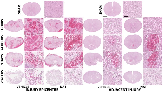
Figure 3. H&E staining demonstrates the morphological changes within the injury epicentre from 5 hours to 2 weeks post-SCI.
Hemorrhage was predominant within 24-SCI, with marked tissue loss observed by 2 weeks post-SCI. No differences were observed between vehicle and NAT treatment groups. Cross section scale bar = 1 mm, high magnification scale bar = 100 µm.

