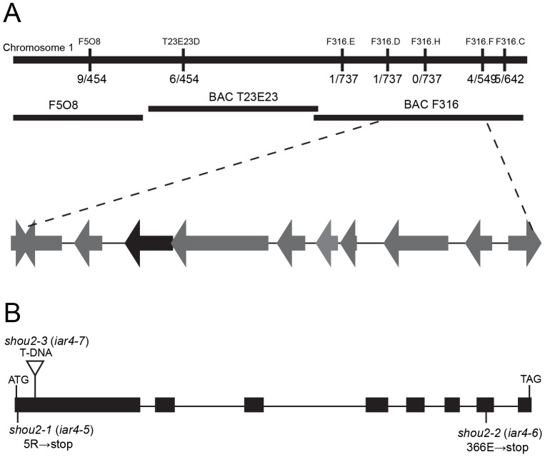
Figure 2. Positional cloning of SHOU2.
(A) shou2 was mapped to a region on chromosome 1 between markers F3I6.D and F3I6.F as described in the methods. The name of each DNA marker is shown above and the number of recombinants is indicated below the line. Open reading frames located between markers F3I6.D and F3I6.F are shown below BAC F316. (B) Structure of SHOU2 gene. Boxes represent exons and lines represent the introns. The positions and changes of the three shou alleles are indicated. The triangle indicates the position of the DNA insertion in shou2-3. The corresponding allele numbers for the iar4 designations are shown in parentheses.

