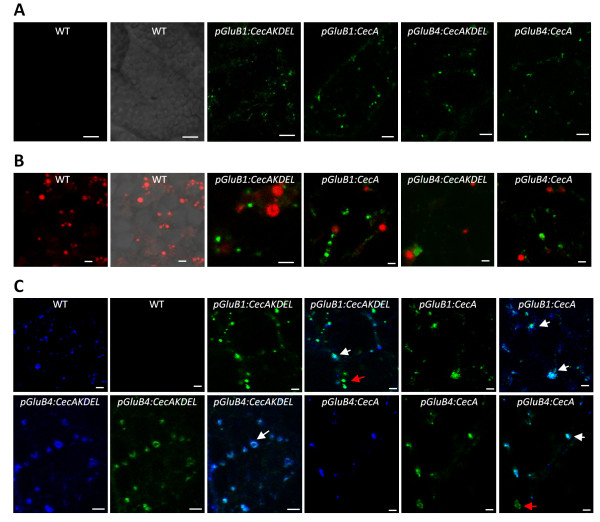
Figure 4.

Cecropin A accumulates in type II protein bodies. Confocal fluorescent images of peripheral endosperm cells from wild-type (WT) or transgenic seeds carrying the transgenes indicated. Cecropin A was immunodetected and visualized in green using an AlexaFluor488-conjugated secondary antibody. Prolamine-containing PBs (PB-I marker) were stained with rhodamine B hexyl ester and visualized in red. Glutelin-containing PBs (PB-II marker) were immunodetected using AlexaFluor647-conjugated anti-glutelin antibodies and visualized in blue. Sections were single fluorescent labelled for cecropin A (A) or double fluorescent labelled for cecropin A and prolamines (B) or for cecropin A and glutelins (C). Images correspond to sequential scan single slides. Merged images are shown in B and C. Scale bars correspond to 10 μm (A) or 2 μm (B and C). Red arrows indicate single immunolabelled vesicles and white arrows double immunolabelled vesicles.
