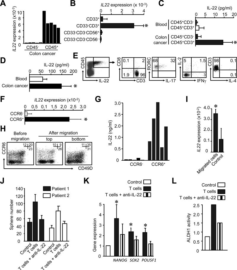
Figure 2. IL-22+ CD4+ T cells traffic into the tumor via CCR6-CCL20 and promote cancer stemenss via IL-22.
(A, B) Different cell populations were sorted from colon cancer tissues and IL-22 expression was measured by real time PCR. (5-10 donors, P < 0.05).
(C) Different CD45+ immune subsets (106/ml) were sorted from colon cancer and blood, and cultured for 12 hours. IL-22 was detected via ELISA. (n = 5, *P < 0.05).
(D) CD45+ subsets (106/ml) were sorted from colon cancer and blood, and were activated with anti-CD3 and anti-CD28 for 2 days. IL-22 was detected by ELISA (n = 5, *P < 0.05).
(E) Single colon cancer environmental cells were stained with anti-CD3, CD8, CD45, RORγc, IL-2, IL-4, IL-17, IL-22 and IFNγ antibodies. The phenotype and the expression of indicated cytokines were analyzed by FACS. Right panels were gated on IL-22+CD45+ cells. One of 3 independent experiments is shown.
(F, G) CD4+ T cells were sorted based on CCR6 expression and activated with anti-CD3 and anti-CD28 and antigen presenting cells. IL-22 mRNA was detected by real time PCR (F) and IL-22 protein by ELISA (G). (5 different donors, P < 0.05)
(H, I) Migration assay was conducted with CD4+ T cells for 4 hours in the presence of CCL20. The phenotype of migrated cells, non-migrated cells, and control (before migration) was analyzed by FACS (H). IL-22 expression was quantified with real-time PCR in the migrated and non-migrated cells. Results are expressed as the mean relative expression (I). (n = 3, P < 0.05).
(J) Sphere assay was performed with autologous colon tumor cells in the presence of activated colon cancer-associated T cells in a transwell system. Anti-IL-22 or isotype mAb was added in the assay. Results are shown as the mean numbers of spheres in triplicates. (2 of 5 patients are shown. P < 0.01).
(K, L) Primary colon cancer associated T cells were sorted and activated for 3 days. DLD-1 colon cancer cells were cultured with these T cell supernatants in the presence of anti-IL-22 or isotype mAbs. The mRNA of stem cell core genes was detected by real-time PCR after 6 hours (K) and ALDH activity was detected by FACS after 48 hours (L). (n = 5, *P < 0.05, compared to control and T cells with anti-IL-22).

