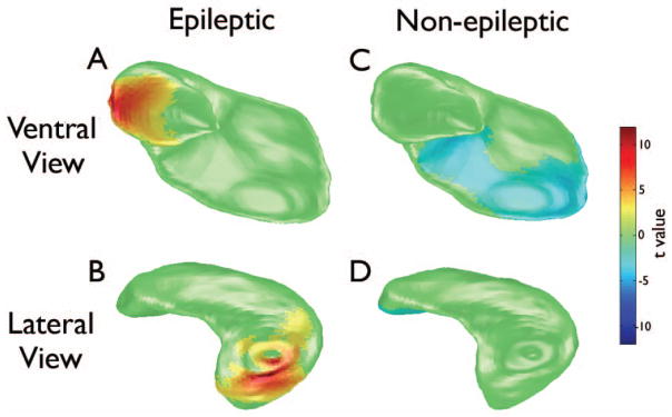
Figure 3.

Large-deformation high-dimensional mapping (HDM-LD) of the ipsilateral hippocampus illustrating changes in epileptic (A and B) and non-epileptic rats (C and D) one week after LFPI compared to baseline. Epileptic rats displayed significantly thicker surface in the lateral region of the hippocampus at one-week post-LFPI (red-yellow) compared to baseline (unpaired two-tailed t test, p < .05, corrected for multiple comparisons). Non-epileptic rats displayed significantly thinner surface in the medial-ventral region at one-week post-LFPI (blue) compared to baseline.
