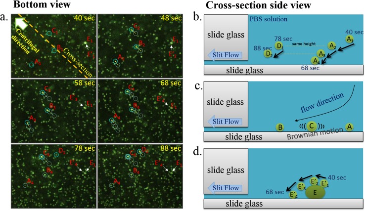
Figure 5.
(a) Snapshots from bottom-view videos from 40 s to 88 s after the PBS solution containing cells were loaded onto the chip. (b) As a cell sinks from the top to bottom of the well, its lateral velocity deceases. Cell D at 78 s is about the same height as cell A at 48 s, and their lateral movement is about the same over 10 s. (c) As shown in cell C, some cells may still move slightly after they settle at the bottom of the well due to Brownian movement. This phenomenon provides an additional mechanism for tighter cell organization. (d) The movement of the E and E′ bright spot between 40 and 68 s shows that cells stacked on top of other cells will continue to move in fluidic fields and find gaps or voids among lower cells. This is consistent with numerical analysis. This phenomenon shows why the cells do not stack and ultimately form tight organization on the chip.

