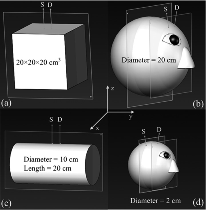
Figure 1.
Four tissue models with different volumes and geometries: (a) a large slab representative of large flat tissue, (b) a large sphere representative of human head, (c) a large cylinder representative of human lower leg, and (d) a small sphere representative of mouse head. The separations between the source (S) and detector (D) fibers were 2.5 cm for the models of (a), (b), and (c), and 0.8 cm for that of (d). The S-D fibers were placed in the ways to mimic in vivo experimental configurations. Note that the illustration scales may not be the same among different models.

