
Figure 1.
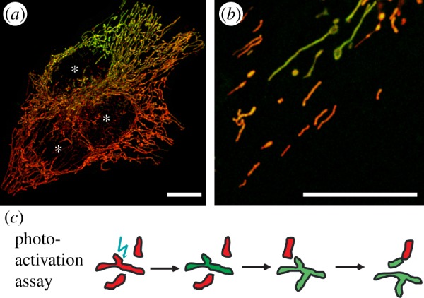
Assays to determine the exchange rates of compounds between mitochondria. (a) Polyethylene glycol- (PEG-)-mediated fusion of cells with differently fluorescent-tagged compounds, here OXPHOS complexes in red (RFP-tagged) and green (GFP-tagged). (b) Use of photo-activatable fluorescent proteins, here OXPHOS complex CI-paGFP to monitor spreading in MitoTracker stained mitochondria. (c) Scheme of photo-activation assay: paGFP is photo-activated in single mitochondria that express paGFP-tagged proteins. Owing to ongoing fusion and fission and mixing of compounds, the fluorescent signal spreads throughout the mitochondrial reticulum. Asterisks indicate nuclei. Scale bars, 10 µm. Images from K.B. (Online version in colour.)