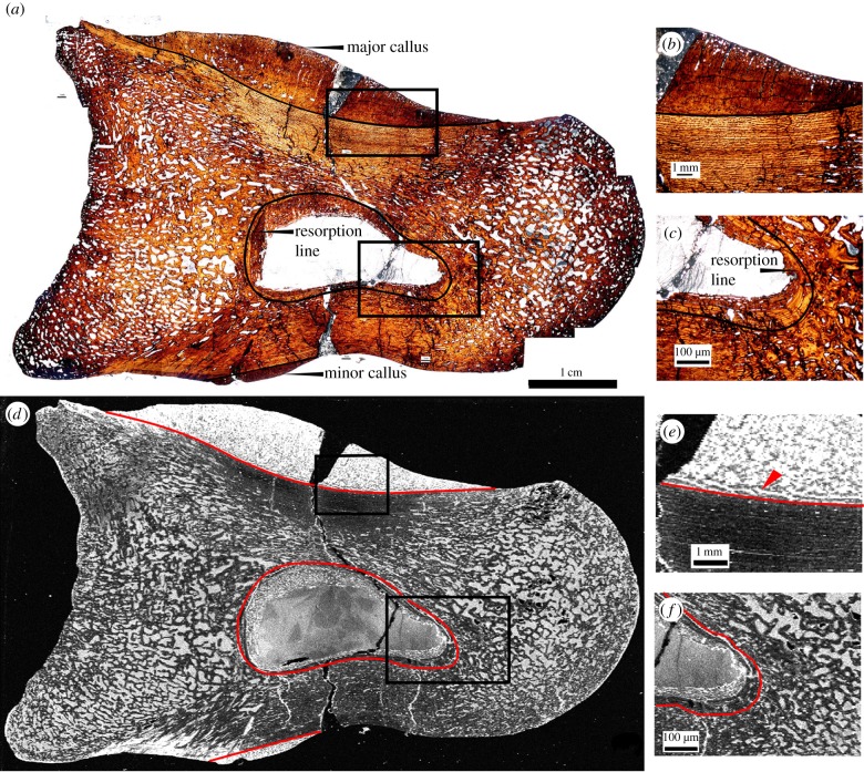
Figure 1.
Thin section of Allosaurus fragilis (specimen UMNH 6282) pedal phalanx as seen in optical observation under plain polarized light (a–c) and in an elemental map of iron taken at the SSRL beamline 6-2 (d–f; grey scale relative image with white as high concentration, black as low). Calli are present on the dorsal (major callus) and ventral (minor callus) surfaces in both optical and elemental images. Photomicrographs (b,c,e,f) represent magnified views of areas of interests (boxed areas), including the major callus (b,e) and resorption cavity (c,f). Laminar orientation of bone tissue is observed in the map of iron around the interface of the major callus and normal cortical bone (e) that is not seen in thin section (b). The extent of resorption and woven bone infill of the medullary cavity are also enhanced in iron (f) compared with thin section (c).

