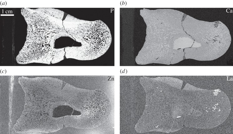
Figure 3.
Elemental maps of phosphorus (a), calcium (b), zinc (c) and lanthanum (d) in UMNH 6282 taken at the SSRL beamline 6-2. Phosphorus is concentrated within the original tissue (a), whereas calcium concentrations are uniform throughout the entire specimen excluding the glass mounting slide of UMNH 6282 (b). Lanthanum is enriched in the pathological tissues of the calli (d). Owing to spectral overlap, the map of lanthanum convolves both lanthanum and barium. Lanthanum was distinguished from barium using PyMCA analysis [44].

