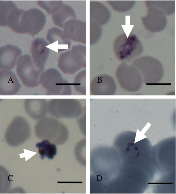
Figure 1.

Parasites images (arrow heads) from Pv positive (panels A-C) and Pf positive (panel D) slides. Panel A shows a young trophozoite or late ring stage parasite in an intact red cell. Panel B shows a late stage P. vivax (Pv) trophozoite in an intact red blood cell. Panel C shows a schizont out of the red cell that does not look healthy. Panel D shows a typical P. falciparum (Pf) ring-stage parasite with double chromatin dots and pale blue cytoplasm. Scale bars = 10 μm).
