
Figure 2.
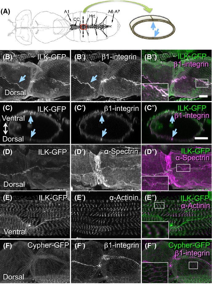
Integrin-linked kinase (ILK) and β1-integrin are localized to cell–cell contact sites and Z-disks in fly hearts. (A) Left, a schematic diagram of the adult fly heart (adopted from Cammarato et al., 2008). Hrt, fly heart tube; CC, conical chamber; A1, abdominal segment 1; A6-A7, abdominal segments 6 and 7. The region outlined in red corresponds to the area shown in B, D, E, and F. Right, a schematic diagram of a cross section of the heart tube along the dotted line in the left panel. The circumference of the heart tube is composed of two myocardial cells. Myocardial cell–cell contact sites are pointed out by arrows. (B-B”) Dorsal views of the heart tube. Both ILK (B and green in B”) and β1-integrin (B’ and magenta in B”) are expressed in adult hearts and colocalize. (C-C”) Optical cross sections from B-B” show circumference of the heart tube. The ventral side is to up. Prominent signals of ILK and β1-integrin are indicated by arrows in B–C”. (D-D”) ILK (D and green in D”) was found at the cell membrane labeled with α-spectrin (D’ and magenta in D”). (E-E”) Ventral views of the heart tube. ILK (E and green in E”) was also localized to Z-disks marked by α-actinin (E’ and magenta in E”). (F-F”) Dorsal views of the heart tube. β1-integrin (F and magenta in F”) is also associated with Z-disks marked by cypher-GFP (F’ and green in F”). All the images in B–F” are in abdominal segment 3 region. Insets in B”, D”, E”, and F” are higher magnification images corresponding to the each rectangle in the respective figures. Bars: 20 μm.