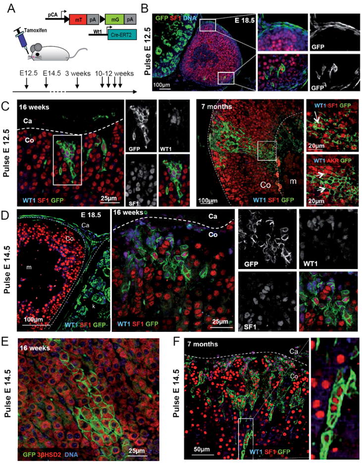
Fig. 4. WT1 expressing cells are progenitors able to differentiate into steroidogenic cells.

(A) Schematic representation of the cell lineage tracing experiments performed on Wt1:Cre-ERT2; mTmG mice or embryos indicating the time point when tamoxifen was administered. (B-F) WT1 or DNA (blue), SF1, 3βHSD2 or AKR1b7 (red) and GFP (green) immunofluorescence on samples of adrenal glands from Wt1:Cre-ERT2; mTmG mice of different ages treated with tamoxifen at E12.5 (B and C) or E14.5 (D to F). During development the majority of GFP+ cells are localized within the capsule (B and D E18.5). Postnatal patches of WT1+, GFP+ cells expand under the adrenal capsule and few cells acquire a steroidogenic phenotype. F) Quantification of the number of GFP+ patches of cells within the adrenal cortex of E18.5 embryos and 3 weeks old Wt1:Cre-ERT2; mTmG mice treated with tamoxifen at E14.5. The overall number of GFP+ clusters increases from 4.9 ± 2.42 (2.6 ± 1.84 WT1+, 2.3 ± 2.45 WT1-) (N=10) to 16.33 ± 4.78 (1.25 ± 0.84 WT1+, 14.0 ± 4.55 WT1-) (N=5). Co, adrenal cortex; Ca, adrenal capsule; m, adrenal medulla. See also Fig. S4.
