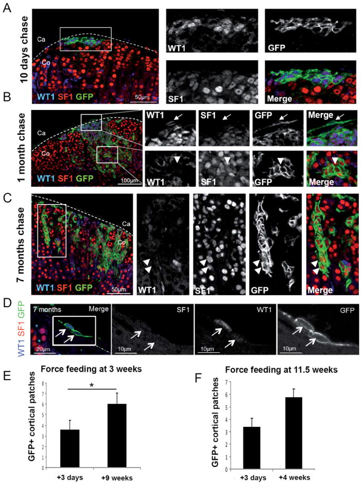
Fig. 5. WT1+ cells maintain the ability to generate subcapsular patches and to differentiate into steroidogenic cells throughout life.

(A-D) Immunostaining against WT1 (blue), SF1 (red) and GFP (green) on adrenals from Wt1:Cre-ERT2; mTmG mice treated with 4 consecutive doses of tamoxifen between 10 and 12 weeks of age and collected after 10 days (A), 1 month (B) and 7 months (C and D) after the last administration. Almost all GFP+ cells also express WT1 and SF1 (A-C). GFP+ SF1+ WT1- cells can be detected in a subset of patches (B-C). GFP+ WT1+ SF1- capsular cells can be detected within the adrenal capsule up to 7 month after force-feeding (D). (E-F) Quantification of the number of GFP+ patches within the adrenal cortex of Wt1:Cre-ERT2; mTmG mice treated with tamoxifen at 3 weeks (E) or 11.5 weeks (F) of age. The number of GFP+ clusters increases significantly from 3.56 ± 2.70 (N=9) to 6.00 ± 2.35 (N=9), 3 days and 9 weeks after force-feeding at 3 weeks of age, respectively. No significant increase in the number of GFP+ clusters is detected in animals force-fed at 11.5 weeks, from 3.38 ± 2.00 (N=8) to 5.75 ± 1.98 (N=8). Co, adrenal cortex; Ca, adrenal capsule. See also Fig. S5.
