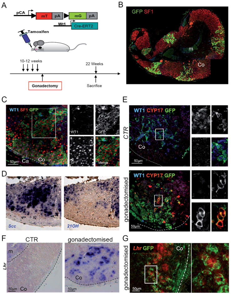
Fig. 6. WT1+ cells generate cells with gonadal characteristics in the adrenal cortex upon gonadectomy.

(A) Schematic representation of the experimental design. (B-E) Adrenal glands from mice sacrificed 10 weeks after gonadectomy. Gonadectomy induces massive invasion of the adrenal cortex by GFP+ cells, derived from WT1+ cells (B). Higher magnification shows that the majority of GFP+ cells have lost WT1 expression, whereas SF1 is present (C). (D) RNA in situ hybridization shows that cells expressing cytochrome P450scc, do not express the adrenocortical specific marker 21-hydroxylase. Please note that the three stainings in panels C to D were performed on consecutive sections, thus representing the same area. (E) CYP17 (red), a marker of cells producing sex hormones (gonadal cells) appears in some cells within the adrenal cortex of gonadectomised mice, but is never found in control animals. Coexpression of CYP17 and GFP in the same cells indicates that they are directly derived from WT1 expressing cells. (F-G) LH receptor RNA in situ hybridization shows expression of this gene in gonadectomised animals (F), where it colocalises with GFP+ cells (G). Co, adrenal cortex; Ca, adrenal capsule; m, adrenal medulla. See also Fig. S6.
