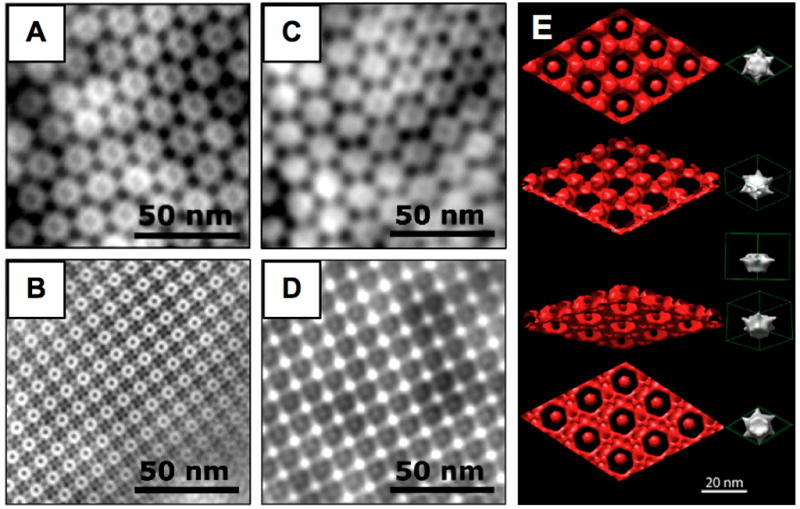
Fig. 1.

TEM images of negatively stained D. radiodurans (A) and S ureae (B) S-layers and of the corresponding electrodeposited Cu2O films (C,D). (E) TEM-based 3D reconstruction of nanostructured Cu2O (red). Four different angles are shown along with a protein unit cell (right). Panel E is reprinted with permission from reference [15].
