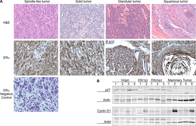
Figure 2. Histopathological characterization of MMTV-Espl1 tumors.
(A) Four histological subtypes (spindle-like, solid, glandular, and squamous) of MMTV-Espl1 multiparous mouse mammary tumors were observed. Representative Hematoxylin and eosin (H&E) stained images (top panel) and ERα positive nuclear and cytoplasmic staining (middle panel) is shown. A spindle-like tumor section is shown for the negative staining using Rabbit IgG and secondary antibody treatment (bottom panel) indicating the absence of positive, brown nuclear and cytoplasmic staining specific for ERα (B) Western blot showing loss of cyclin dependent kinase (CDK) inhibitor p27 expression in 4 out of 5, and higher expression of Cyclin D1 in 2 out of 5 of mammary tumors compared to multiparous mammary glands. β-Actin is shown to compare loading.

