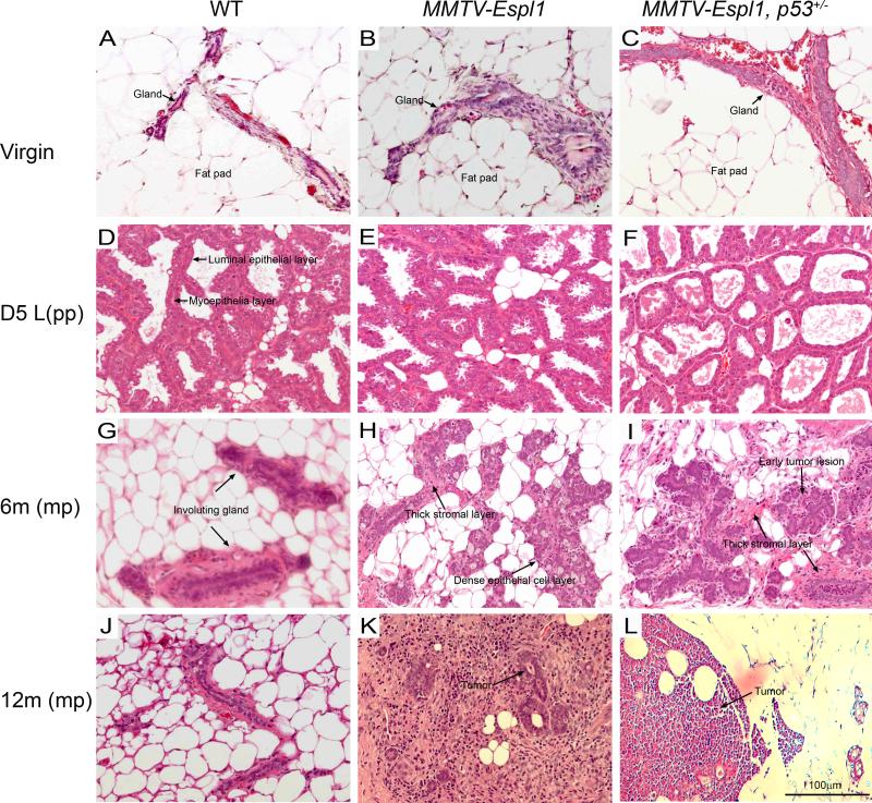
Figure 3. Histological characterization and progression of mammary tumorigenesis in MMTV-Espl1 transgenic mammary glands.
Mammary morphologies in Hematoxylin and eosin (H&E)-stained sections of Virgin (A, B, C) , Day five (D5) lactation (D, E, F), D5 post force weaned at D1, 6 month old mice (G, H, I) and D5 post force weaned at D1, 12month old mice (J, K, L) of wild type (WT), MMTV-Espl1 and MMTV-Espl1, p53+/− genotype are shown. Mammary glands from virgin WT, MMTV-Espl1 and MMTV-Espl1, p53+/− mice display a normally sparse and isolated mammary gland epithelium within large areas of mammary fat pad. MMTV-Espl1 and MMTV-Espl1, p53+/− mice also display normal looking mammary gland architecture at day 5 lactation (E, F) with regular luminal epithelial ductal structure and a thin myoepithelial layer surrounding the luminal cells comparable to WT littermate mice (D, arrows). The epithelial cells in multi-pregnant 6 month old MMTV-Espl1 mice at day five post day one force weaned mice display dense epithelial cell clusters (H, I, arrows) and thick stromal layers (H, I, arrows) compared to WT litter mate controls that display sparse mammary epithelial cells and involuting mammary glands at this stage (G). Mammary duct hyperplasia and early tumor lesions also develop in 6 month old multiparous MMTV-Espl1, p53+/− mice (I, arrows). At 12 months post day one lactation 80% MMTV-Espl1 mice and 100% MMTV-Espl1, p53+/− mice show mammary tumor development with palpable mammary tumor formation (K, L, arrows). (n = 3 for each genotype. Representative images are shown for all animals.)

