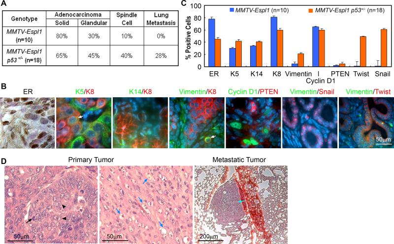
Figure 4. MMTV-Espl1 transgenic mice have highly heterogeneous ERα positive mammary tumors with variable markers for luminal and basal epithelial cells.
(A) Occurrence of each tumor morphology/feature for all tumors within a genotypic class (n = number of tumors; p=0.0633, Fisher's exact test). (B) Representative immunohistochemical and immunofluorescence staining for ERα brown), Keratin-5 (K5, green), Keratin-8 (K8, red), Keratin-14 (K14, green), Vimentin (green), Cyclin D1 (green), PTEN (red), Snail and Twist (red). Keratin-8 (green) and Keratin-5 (red) label luminal and myoepithelial cells, respectively, with regions of co-expressing cells (arrow). Metaplastic tumor cells show dual staining of Keratin-8 and the mesenchymal marker, Vimentin (arrow). (C) The quantification (% positive cells) for all markers, (p<0.0001) from 10 MMTV-Espl1 and 18 MMTV-Espl1 p53+/− tumors. Approximate 100 cells from each tumor were analyzed. (D) Solid morphology of the primary tumors, with high levels of mitosis (arrows) and pleomorphic nuclei (arrowheads). Homogeneous, fusiform, spindloid cells of a primary carcinosarcoma type mammary tumor entrap carcinomatous cells are shown (blue arrows). Pulmonary metastases (cyan arrow) were observed in MMTV-Espl1, p53+/− tumors. Because the tumors showed mixed morphological phenotypes, regardless of genotype, the occurrence of each morphology/feature for all tumors within a genotypic class was tabulated (A, C).

