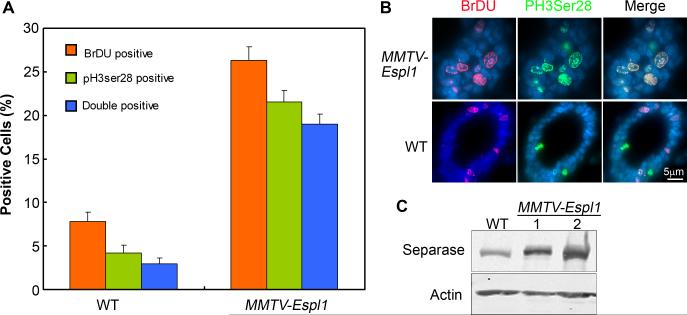
Figure 8. MMTV-Espl1 mice display early cell cycle defects and co-localization of BrdU positive (S phase) and PH3 (Ser28, mitotic phase) cells.
(A-B) Mid-pregnant mammary gland sections were stained for BrdU (red fluorescence) and Phospho Histone H3 (Serine 28, green fluorescence). Representative images are shown for MMTV-Espl1 and wild type (WT) mice (B) and quantified in (A) as a percentage of total cells positive for DAPI (p<0.0005). (C) Western blot shows increased Separase expression in MMTV-Espl1 mice mid-pregnant glands compared to WT. (n=3 animals used for each genotype, and ~200 cells per animal were analyzed).

