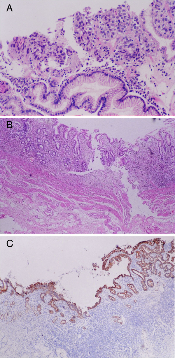
Figure 1.

Tumor and histopathological features. (A) Biopsy indicated undifferentiated adenocarcinoma. (B) Microscopic examination of the excised specimen showed no remaining gastric adenocarcinoma cells and coverage of the lesion with regenerative mucosa. Gland degeneration was seen in the mucosa. Fibrosis and lymph follicle formation were observed in the gastric wall. (C) No gastric adenocarcinoma cell remnants were detected even with immunohistochemistry staining for CK20.
