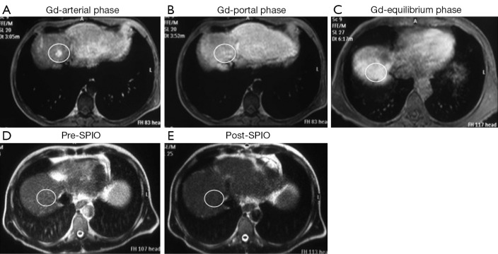
Figure 2.
A small (1 cm) dysplastic nodule, proven by biopsy, was located in the segment VIII; dynamic magnetic resonance (MR) images after gadolinium (Gd) administration show focal faint enhancement by the lesion (white circle) only in the arterial phase (A) with no focal abnormality in portal (B) and equilibrium (C) phases (score =1; false positive finding); T2 weighted MR images before (D) and after (E) superparamagnetic iron oxide (SPIO) administration show homogeneous contrast distribution in the liver with diffuse hypointensity and no focal abnormalities (score =0; true negative finding).

