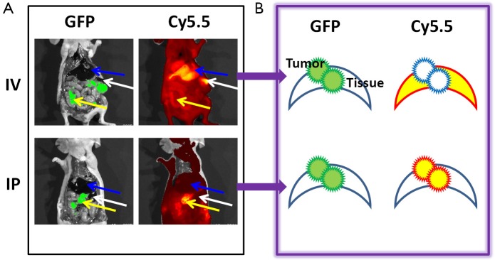
Figure 1.

Fluorescence images of ovarian tumors with ProSense (2 nmoles) delivered through IV or IP injection 24 h before imaging. (A) GFP signal indicated the location of the SKOV3 tumor, and Cy5.5 images showed the protease-activated ProSense signal. IV injection of the probe resulted in high Cy5.5 signals in the liver (blue arrow) and spleen (white arrow) but not in small tumors (yellow arrow). In contrast, IP injection of the probe resulted in high Cy5.5 signals in tumors and low signals in the liver and spleen; (B) Schematic drawing of the fluorescence contrast between tumors and organs according to the route through which the probe is administered. GFP, green fluorescent protein; IV, intravenous; IP, intraperitoneal.
