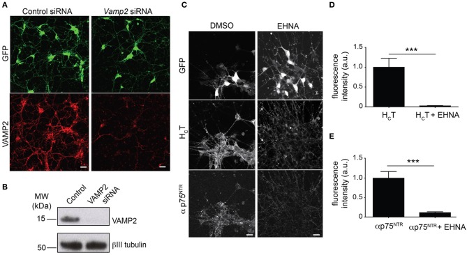
Figure 1.
Optimization of the read-out for the siRNA screen. (A) Representative images of GFP-positive HBG3 ES cell-derived motor neurons (green) reverse transfected with Vamp2 siRNA using Dreamfect Gold in a 96-well plate format and immunostained for VAMP2 (red). Scale bar, 20 μm. (B) Western blot analysis of VAMP2 after siRNA mediated Vamp2 knock-down in HBG3 ES cell-derived motor neurons transfected in a 96-well format. (C) HBG3 ES cell-derived motor neurons expressing GFP (upper panels) were incubated with AlexaFluor555-conjugated HCT (middle panels) and αp75NTR (lower panels) in the presence of 1 mM EHNA or DMSO vehicle control for 2 h at 37°C. Cells were then acid-washed, fixed and immunostained with AlexaFluor488-conjugated anti-rabbit IgG. The intracellular accumulation of both probes was substantially reduced by EHNA treatment, indicating that cytoplasmic dynein plays a major role in this process. Scale bar, 20 μm. (D,E) Quantification of internalized HCT (D) and αp75NTR (E) in motor neurons exposed to HCT and αp75NTR from three independent experiments with or without EHNA (paired t-test, mean ± s.e.m., ***p = 0.001).

