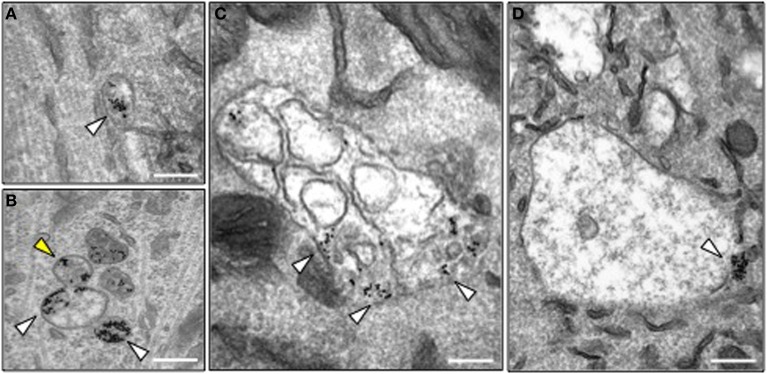
Figure 4.

Morphology of HCT-containing organelles in wild type and Bicd1gt/gt motor neurons. (A) Transmission electron microscopy image of a typical endosome containing gold-conjugated HCT (white arrowhead) in wild type motor neurons. HCT was internalized for 2 h at 37°C. Scale bar, 200 nm. (B–D) Transmission electron microscopy images of representative organelles containing gold-conjugated HCT (white arrowheads) in Bicd1gt/gt motor neurons following internalization for 2 h at 37°C. HCT was often detected in budding structures (B, yellow arrowhead), or enlarged endosomal organelles containing membranes (C) or with an amorphous content (D). In several instances, gold-conjugated HCT was found clustered at the limiting membrane of these structures. Scale bar, 200 nm.
