Figure 5.
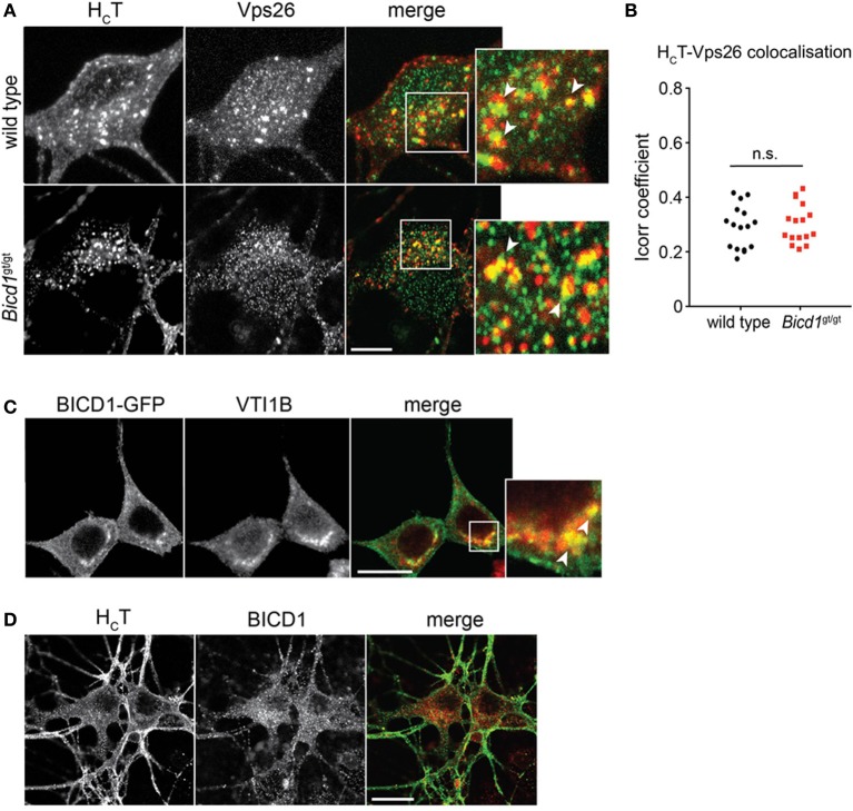
HCT partially co-distributes with retromer components and late endosomal SNAREs. (A) Wild type and Bicd1gt/gt motor neurons were incubated with AlexaFluor555-conjugated HCT (red) for 1 h at 37°C, then acid-washed, fixed and immunostained for Vps26 (green). Insets from the merged images are magnified to show co-localization (yellow) between HCT and Vps26. Scale bar, 5 μm. (B) Quantification of HCT/Vps26 co-localization from the experiment shown in (A) (16 cells per condition, Mann-Whitney test, mean ± s.e.m., n.s. non-significant). (C) N2A cells over-expressing BICD1-GFP (green) were fixed and immunostained for VTI1B (red). The white box in the merged channel is magnified to highlight the extent of co-localization of BICD1-GFP and this late endosomal SNARE protein. Scale bar, 20 μm. (D) Wild type motor neurons were incubated with AlexaFluor488-conjugated HCT (green) for 1 h at 37°C, acid-washed, fixed and then immunostained for BICD1 (red). Co-localization between HCT and BICD1 was not detected under these conditions. Scale bar, 20 μm.

