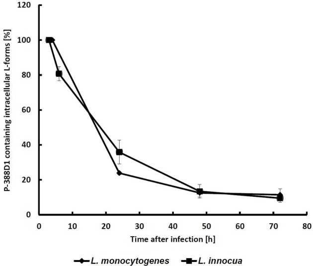
Figure 2.

Monitoring of GFP-labeled intracellular L-forms (ICL) in P-388D1 macrophages. After 3 h p.i. approx. 1.2% of the macrophage population featured intracellular L-forms, which was set as the initial 100% value. Fate of the ICL was monitored for 72 h. Values represent the means of five microscopic fields in two replicate experiments. Standard deviation is indicated by error bars.
