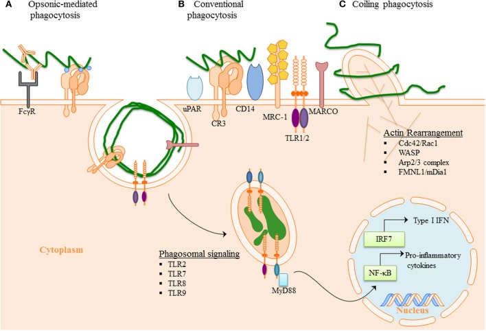
Figure 2.
Phagocytosis of B. burgdorferi: (A) Opsonic-mediated phagocytosis—complement factors, such as C3b and iC3b, bound to the surface of Bb can interact with complement receptors and mediate phagocytosis. Additionally, the immune cells Fc-receptors have the ability to bind to the opsonic antibodies that coat Bb and internalize the pathogen. (B) Conventional phagocytosis—the direct interaction of surface receptors with Bb, such as integrins and C-type lectins, allows for tether of the spirochete to the cell surface. Various PRRs induced signal cascade initiates formation of the phagocytic cup and spirochete engulfment. (C) Coiling phagocytosis—the preferred mechanism of spirochete internalization in which the phagocytic cell uses filopodial protrusions that capture Bb. The filopodial enwrap the spirochete and convert into coiling pseudopods. During this dynamic process FMNL1, mDai1 Arp2/3 complex, and WASP are involved in actin rearrangement of the cell, which then facilitates subsequent phagocytosis of Bb. Following internalization of Bb, the spirochete is degraded within the phagosome thus exposing additional PAMPs to PRR with in the phagosome. The phagosomal signals initiated by Bb generates a robust inflammatory response, including the induction of pro-inflammatory genes and Type I IFNs.

