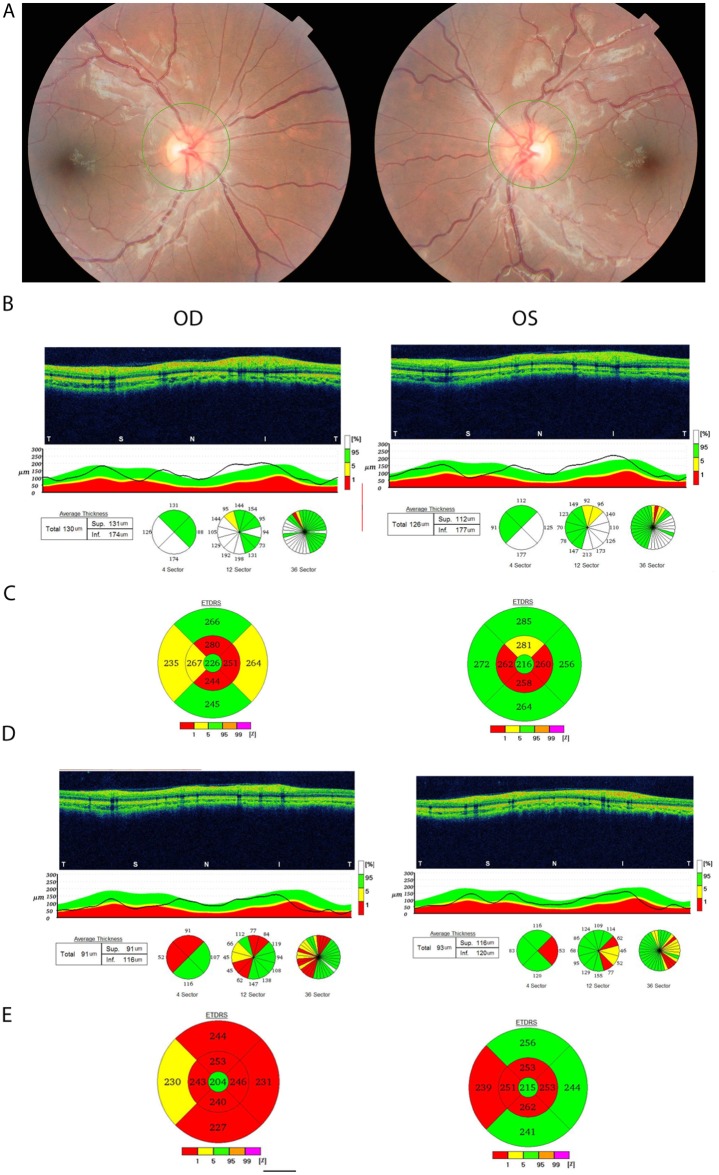
Figure 2 .
(A) Fundus photograph of proband VI-9 showing mild bilateral swelling of optic nerves. (B) Optical coherence tomography (OCT) of the retinal nerve fibre layer showing mild thickening of the inferior and temporal quadrants with resolution and subsequent thinning 8 months later (D). (C) OCT of the macula showing reduced macular thickness in both eyes and further thinning 8 months later (E). OD, right eye; OS, left eye.

