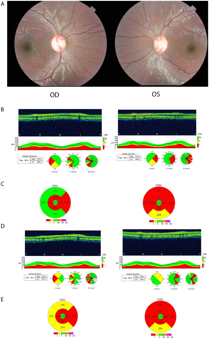
Figure 3 .
(A) Fundus photograph of proband VI-19 showing optic nerve head pallor in both eyes. (B) Optical coherence tomography (OCT) of the retinal nerve fibre layer (RNFL) shows thinning in the temporal quadrant and repeat OCT 6 months later (3D) shows further thinning of the RNFL extending into the superior and inferior quadrants. (C) OCT of the macula shows thinning and a repeat OCT 6 months later shows progressive thinning as well (E). Four other family participants (VI-10, VI-17, VI-20 and V-1) received full clinical assessment and showed similar features (data not shown). OD, right eye; OS, left eye.

