
Figure 1.
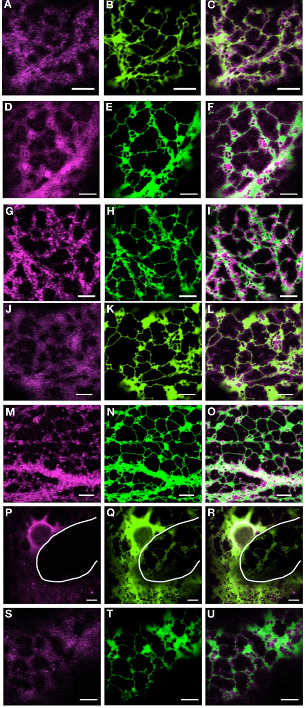
Coexpression of myosin tail domains with an ER marker. Representative images of cells coexpressing GFP-HDEL (green) and mRFP myosin tail domain fusions [magenta; XI-A, (A–C); XI-1, (D–F); XI-2, (G–I); XI-C, (J–L); XI-E, (M–O); XI-I, (P–R); XI-K, (S–U)]. White line in panels (P–R) highlights the cell boundary between neighboring cells. Scale bar 5 μm.