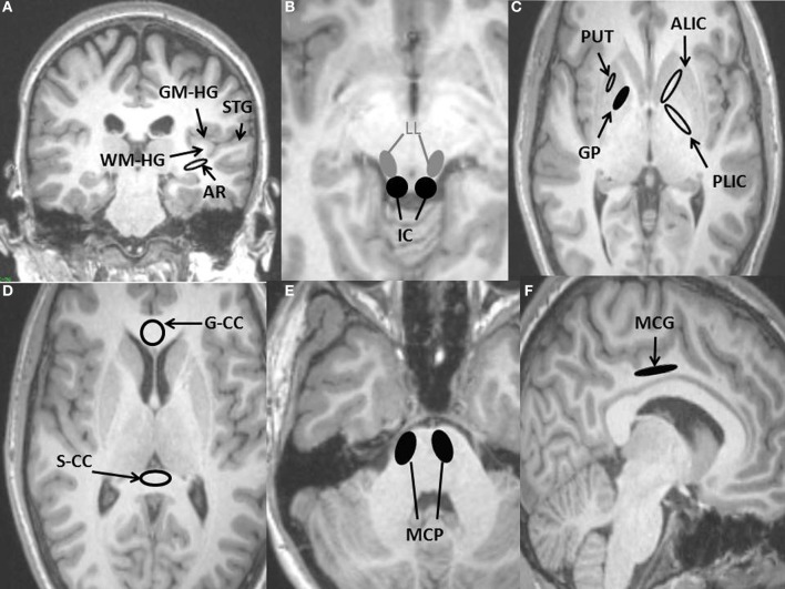
Figure 1.
Regions of interest (ROIs) used for DTI measurements placed on anatomical T1-weighted MRI scan. (A) Four auditory ROIs, Gray matter of Heschl's gyrus (GM-HG), White matter of Heschl's gyrus (WM-HG), Superior temporal gyrus (STG), and Auditory radiation (AR). (B) Two auditory ROIs, Inferior colliculus (IC), Lateral lemniscus (LL). (C) Three non-auditory ROIs, Putamen (PUT), Globus pallidus (GP), Posterior limb of the internal capsule (PLIC), Anterior limb of internal capsule (ALIC). (D) Two non-auditory ROIs, Genu of the corpus callosum (G-CC), Splenium of the corpus callosum (S-CC). (E) Non-auditory ROI, Middle cerebellar peduncle (MCP). (F) Non-auditory ROI, Middle cingulate gyrus (MCG).

