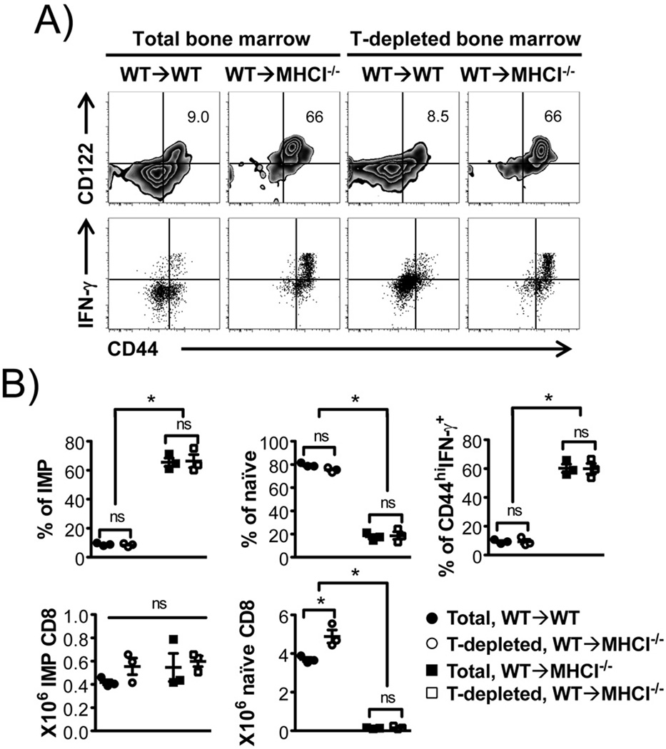
FIGURE 2. IMP CD8+ T cells in WT→MHCI−/− chimeras develop despite depletion of T cells from donor bone marrow.
WT bone marrow was either left intact or depleted of T-cells, and used as donors to generate “WT→WT” and “WT→MHCI−/−” chimeras. Donor TCRβ+CD8α+ cells were analyzed. (A) Flow cytometric analysis for CD44 and CD122, and percentage of CD44hi IFN-γ producing CD8+ T cells in response to P/I. (B) Percentages and numbers of IMP and naïve CD8+ T cells, and percentage of P/I induced CD44hi IFN-γ+ CD8+ T cells. n = 3 in each group. *p < 0.05, ns = not significant, by Student’s t test.

