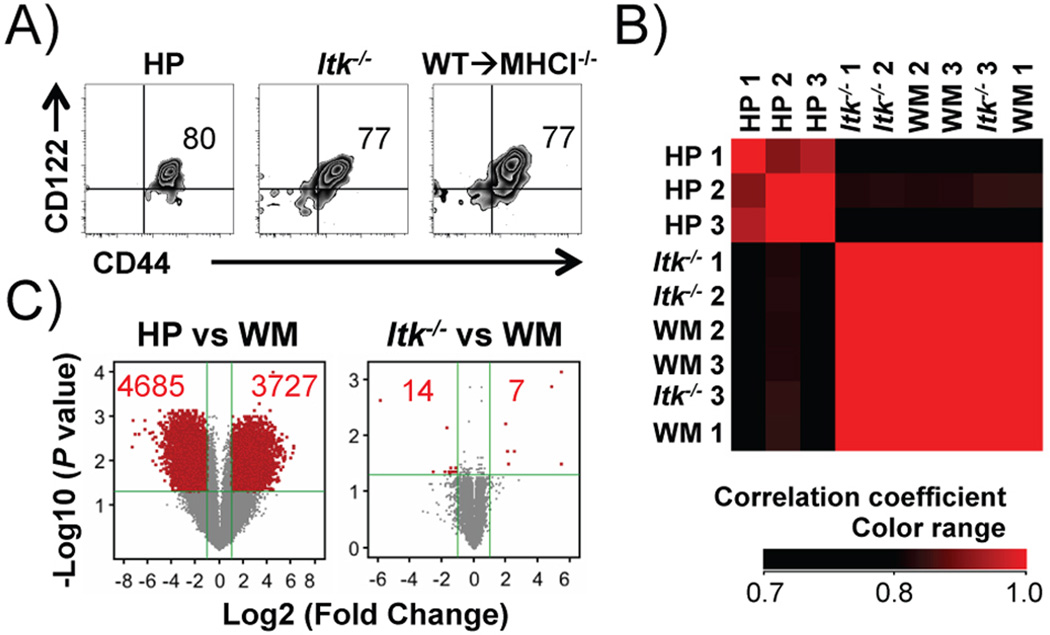
FIGURE 3. Hematopoietic MHCI dependent CD8+ T cells resemble innate memory CD8+ T cells in Itk−/− mice, but are distinct from HP cells.
Comparison of HP, Itk−/− and WM (WT→MHCI−/−) CD8+ T cells. (A) HP, Itk−/− and WM CD8+ T cells share expression of CD44 and CD122. (B) Itk−/− and WM CD8+ T cells show extremely high correlation in whole genome gene expression, which is distinct from HP CD8+ T cells. Samples were clustered based on the hierarchy of correlation. (C) HP but not Itk−/− CD8+ T cells, exhibit significantly higher number of differentially expressed genes compared to WM CD8+ T cells. Genes with significant change (fold change > 2, corrected P < 0.05) are shown in red. Numbers in red indicate numbers of gene that significantly up- or down- regulated.

