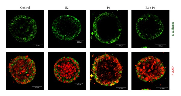
Figure 1.

E-cadherin localization in acinar structures formed by BME-UV1 cells cultured on Matrigel for 12 days in differentiation medium (control), enriched with 17β-estradiol (E2, 1 nM), progesterone (P4, 5 ng/mL), or both (E2 + P4); E-cadherin (epithelial cell marker) was labeled with antibodies conjugated with Alexa Fluor 488 (green fluorescence) and DNA was counterstained with 7AAD (red fluorescence).
